

Which Cell Is More Complex
| Cell junction | |
|---|---|
| Details | |
| Identifiers | |
| Latin | junctiones cellulares |
| TH | H1.00.01.0.00012 |
| FMA | 67394 |
| Anatomical terminology [edit on Wikidata] | |
Cell junctions (or intercellular bridges [1]) are a class of cellular structures consisting of multiprotein complexes that provide contact or adhesion between neighboring cells or between a cell and the extracellular matrix in animals. They also maintain the paracellular barrier of epithelia and control paracellular send. Jail cell junctions are peculiarly abundant in epithelial tissues. Combined with cell adhesion molecules and extracellular matrix, cell junctions help agree fauna cells together.
Cell junctions are likewise particularly of import in enabling communication between neighboring cells via specialized protein complexes chosen communicating (gap) junctions. Jail cell junctions are also important in reducing stress placed upon cells.
In plants, similar advice channels are known as plasmodesmata, and in fungi they are called septal pores.[2]
Types [edit]

Some examples of jail cell junctions
In vertebrates, there are three major types of jail cell junction:
- Adherens junctions, desmosomes and hemidesmosomes (anchoring junctions)
- Gap junctions[3] (communicating junction)
- Tight junctions (occluding junctions)
Invertebrates have several other types of specific junctions, for instance septate junctions or the C. elegans apical junction. In multicellular plants, the structural functions of cell junctions are instead provided for by cell walls. The analogues of communicative cell junctions in plants are chosen plasmodesmata.
Anchoring junctions [edit]
Cells within tissues and organs must be anchored to one some other and attached to components of the extracellular matrix. Cells accept developed several types of junctional complexes to serve these functions, and in each case, anchoring proteins extend through the plasma membrane to link cytoskeletal proteins in one jail cell to cytoskeletal proteins in neighboring cells as well as to proteins in the extracellular matrix.[iv]
3 types of anchoring junctions are observed, and differ from one another in the cytoskeletal poly peptide anchor as well as the transmembrane linker protein that extends through the membrane:
| Junction | Cytoskeletal anchor | Transmembrane linker | Ties jail cell to: |
|---|---|---|---|
| Desmosomes | Intermediate filaments | Cadherin | Other cells |
| Hemidesmosomes | Intermediate filaments | Integrins | EC matrix |
| Adherens junctions (Adhesion belt, Focal adhesion) | Actin filaments | Cadherin / Integrins | Other cells / EC matrix |
Anchoring-type junctions not simply hold cells together only provide tissues with structural cohesion. These junctions are most abundant in tissues that are subject area to abiding mechanical stress such as peel and heart.[four]
Desmosomes [edit]

This prototype shows a desmosome junction between cells of the epidermal layer of the peel.
Desmosomes, too termed as maculae adherentes, can be visualized as rivets through the plasma membrane of adjacent cells. Intermediate filaments composed of keratin or desmin are fastened to membrane-associated attachment proteins that form a dense plaque on the cytoplasmic confront of the membrane. Cadherin molecules form the bodily anchor by attaching to the cytoplasmic plaque, extending through the membrane and binding strongly to cadherins coming through the membrane of the adjacent cell.[5]
Hemidesmosomes [edit]
Hemidesmosomes course rivet-like links betwixt cytoskeleton and extracellular matrix components such as the basal laminae that underlie epithelia. Similar desmosomes, they tie to intermediate filaments in the cytoplasm, but in dissimilarity to desmosomes, their transmembrane anchors are integrins rather than cadherins.[6]
Adherens junctions [edit]
Adherens junctions share the characteristic of anchoring cells through their cytoplasmic actin filaments. Similarly to desmosomes and hemidesmosomes, their transmembrane anchors are equanimous of cadherins in those that ballast to other cells and integrins (focal adhesion) in those that ballast to extracellular matrix. There is considerable morphologic diversity among adherens junctions. Those that necktie cells to one another are seen as isolated streaks or spots, or as bands that completely encircle the cell. The ring-type of adherens junctions is associated with bundles of actin filaments that also encircle the cell just below the plasma membrane. Spot-like adherens junctions called focal adhesions help cells adhere to extracellular matrix. The cytoskeletal actin filaments that necktie into adherens junctions are contractile proteins and in addition to providing an anchoring function, adherens junctions are thought to participate in folding and bending of epithelial prison cell sheets. Thinking of the bands of actin filaments as being similar to 'drawstrings' allows one to envision how contraction of the bands within a grouping of cells would distort the sheet into interesting patterns.[4]
Communicating (gap) junctions [edit]
Communicating junctions, or gap junctions allow for straight chemic communication between side by side cellular cytoplasm through diffusion without contact with the extracellular fluid.[seven] This is possible due to six connexin proteins interacting to form a cylinder with a pore in the center called a connexon.[eight] The connexon complexes stretches beyond the cell membrane and when two next cell connexons collaborate, they form a complete gap junction channel.[vii] [viii] Connexon pores vary in size, polarity and therefore can exist specific depending on the connexin proteins that institute each individual connexon.[7] [8] Whilst variation in gap junction channels do occur, their structure remains relatively standard, and this interaction ensures efficient communication without the escape of molecules or ions to the extracellular fluid.[8]
Gap junctions play vital roles in the human trunk,[9] including their part in the compatible contractile of the heart muscle.[9] They are likewise relevant in signal transfers in the brain, and their absenteeism shows a decreased cell density in the encephalon.[x] Retinal and skin cells are as well dependent on gap junctions in cell differentiation and proliferation.[9] [10]
Tight junctions [edit]
Found in vertebrate epithelia, tight junctions human activity as barriers that regulate the move of water and solutes between epithelial layers. Tight junctions are classified as a paracellular barrier which is divers every bit non having directional discrimination; however, motion of the solute is largely dependent upon size and charge. There is prove to suggest that the structures in which solutes laissez passer through are somewhat like pores.
Physiological pH plays a part in the selectivity of solutes passing through tight junctions with virtually tight junctions existence slightly selective for cations. Tight junctions present in different types of epithelia are selective for solutes of differing size, charge, and polarity.
Proteins [edit]
There have been approximately 40 proteins identified to be involved in tight junctions. These proteins can be classified into four major categories; scaffolding proteins, signalling proteins, regulation proteins, and transmembrane proteins.
Roles [edit]
- Scaffolding proteins – organise the transmembrane proteins, couple transmembrane proteins to other cytoplasmic proteins as well as to actin filaments.
- Signaling proteins – involved in junctions associates, barrier regulation, and gene transcription.
- Regulation proteins – regulate membrane vesicle targeting.
- Transmembrane proteins – including junctional adhesion molecule, occludin, and claudin.
It is believed that claudin is the poly peptide molecule responsible for the selective permeability betwixt epithelial layers.
A three-dimensional image is withal yet to be accomplished and as such specific data most the function of tight junctions is all the same to be determined.
Tricellular junctions [edit]
Tricellular junctions seal epithelia at the corners of three cells. Due to the geometry of 3-cell vertices, the sealing of the cells at these sites requires a specific junctional organization, different from those in bicellular junctions. In vertebrates, components tricellular junctions are tricellulin and lipolysis-stimulated lipoprotein receptors. In invertebrates, the components are gliotactin and anakonda.[11]
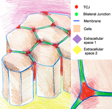
The cartoon of epithelium cells connected by tricellular junctions at the regions where iii cells run across.
Tricellular junctions are likewise implicated in the regulation of cytoskeletal organization and cell divisions. In particular they ensure that cells carve up according to the Hertwig rule. In some Drosophila epithelia, during cell divisions tricellular junctions plant physical contact with spindle apparatus through astral microtubules. Tricellular junctions exert a pulling strength on the spindle apparatus and serve equally a geometrical clues to make up one's mind orientation of jail cell divisions.[12]
Cell junction molecules [edit]
The molecules responsible for creating jail cell junctions include various jail cell adhesion molecules. There are four primary types: selectins, cadherins, integrins, and the immunoglobulin superfamily.[13]
Selectins are prison cell adhesion molecules that play an important role in the initiation of inflammatory processes.[fourteen] The functional capacity of selectin is limited to leukocyte collaborations with vascular endothelium. There are three types of selectins found in humans; L-selectin, P-selectin and E-selectin. Fifty-selectin deals with lymphocytes, monocytes and neutrophils, P-selectin deals with platelets and endothelium and E-selectin deals only with endothelium. They have extracellular regions made upwardly of an amino-terminal lectin domain, attached to a saccharide ligand, growth factor-like domain, and short echo units (numbered circles) that match the complementary bounden poly peptide domains.[xv]
Cadherins are calcium-dependent adhesion molecules. Cadherins are extremely important in the process of morphogenesis – fetal development. Together with an blastoff-beta catenin complex, the cadherin can demark to the microfilaments of the cytoskeleton of the prison cell. This allows for homophilic cell–cell adhesion.[16] The β-catenin–α-catenin linked complex at the adherens junctions allows for the formation of a dynamic link to the actin cytoskeleton.[17]
Integrins deed as adhesion receptors, transporting signals across the plasma membrane in multiple directions. These molecules are an invaluable function of cellular advice, as a single ligand can be used for many integrins. Unfortunately these molecules still have a long fashion to go in the ways of inquiry.[18]
Immunoglobulin superfamily are a group of calcium independent proteins capable of homophilic and heterophilic adhesion. Homophilic adhesion involves the immunoglobulin-like domains on the prison cell surface binding to the immunoglobulin-like domains on an opposing cell's surface while heterophilic adhesion refers to the binding of the immunoglobulin-like domains to integrins and carbohydrates instead.[19]
Cell adhesion is a vital component of the body. Loss of this adhesion furnishings cell structure, cellular functioning and communication with other cells and the extracellular matrix and can pb to severe health bug and diseases.
References [edit]
- ^ Mitchell, Richard Sheppard; Kumar, Vinay; Abbas, Abul G.; Fausto, Nelson (2007). "Ch. xiii: Box on morphology of squamous cell carcinoma". Robbins Basic Pathology (8th ed.). Philadelphia: Saunders. ISBN978-1-4160-2973-1.
- ^ Bloemendal, Due south; Kück, U (January 2013). "Cell-to-cell communication in plants, animals, and fungi: a comparative review". Die Naturwissenschaften. 100 (1): three–xix. Bibcode:2013NW....100....3B. doi:10.1007/s00114-012-0988-z. PMID 23128987. S2CID 11991859.
- ^ Andrew Fifty Harris; Darren Locke (2009). Connexins, A Guide. New York: Springer. p. 574. ISBN978-1-934115-46-6.
- ^ a b c Yan HH, Mruk DD, Lee WM, Cheng CY (2008). Cross-talk between tight and anchoring junctions-lesson from the testis . Advances in Experimental Medicine and Biology. Vol. 636. New York, NY : Springer-Verlag New York. pp. 234–54. doi:10.1007/978-0-387-09597-4_13. ISBN978-0-387-79990-2. PMC4080640. PMID 19856171.
- ^ Lie PP, Cheng CY, Mruk DD (2011). The biological science of the desmosome-like junction a versatile anchoring junction and signal transducer in the seminiferous epithelium. International Review of Cell and Molecular Biology. Vol. 286. pp. 223–69. doi:10.1016/B978-0-12-385859-7.00005-vii. ISBN9780123858597. PMC4381909. PMID 21199783.
- ^ Gipson IK, Spurr-Michaud SJ, Tisdale Equally (April 1988). "Hemidesmosomes and anchoring fibril collagen announced synchronously during development and wound healing". Developmental Biology. 126 (2): 253–62. doi:10.1016/0012-1606(88)90136-four. PMID 3350210.
- ^ a b c Evans WH, Martin PE (2002). "Gap junctions: structure and part (Review)". Molecular Membrane Biology. 19 (two): 121–36. doi:ten.1080/09687680210139839. PMID 12126230. S2CID 20806078.
- ^ a b c d Lampe PD, Lau AF (July 2004). "The effects of connexin phosphorylation on gap junctional advice". International Journal of Biochemistry & Prison cell Biology. 36 (7): 1171–86. doi:10.1016/S1357-2725(03)00264-4. PMC2878204. PMID 15109565.
- ^ a b c "Abstracts: Proceedings of the International Gap Junction Conference. Baronial 5–ix, 2007. Elsinore, Denmark". Cell Communication & Adhesion. xiv (6): 275–346. 2007. doi:10.1080/15419060801891042. PMID 18392995.
- ^ a b Wei CJ, Xu X, Lo CW (2004). "Connexins and cell signaling in development and disease". Almanac Review of Cell and Developmental Biological science. xx: 811–38. doi:10.1146/annurev.cellbio.19.111301.144309. PMID 15473861.
- ^ Byri S, Misra T, Syed ZA, Batz T, Shah J, Boril Fifty, Glashauser J, Aegerter-Wilmsen T, Matzat T, Moussian B, Uv A, Luschnig South (2015). "The triple-repeat protein Anakonda controls epithelial tricellular junction germination in Drosophila". Developmental Prison cell. 33 (v): 535–48. doi:x.1016/j.devcel.2015.03.023. PMID 25982676.
- ^ Bosveld F, Markova O, Guirao B, Martin C, Wang Z, Pierre A, Balakireva Chiliad, Gaugue I, Ainslie A, Christophorou North, Lubensky DK, Minc Northward, Bellaïche Y (2016). "Epithelial tricellular junctions human action equally interphase cell shape sensors to orient mitosis". Nature. 530 (7591): 496–8. Bibcode:2016Natur.530..495B. doi:x.1038/nature16970. PMC5450930. PMID 26886796.
- ^ Lodish; et al. (2007). Molecular Prison cell Biology (6th ed.). W. H. Freeman and Company. p. 803. ISBN978-1429203142.
- ^ Tedder TF, Steeber DA, Chen A, Engel P (July 1995). "The selectins: vascular adhesion molecules". FASEB Periodical. 9 (10): 866–73. doi:10.1096/fasebj.9.ten.7542213. PMID 7542213. S2CID 8315194.
- ^ Bevilacqua MP, Nelson RM (February 1993). "Selectins". Periodical of Clinical Investigation. 91 (two): 379–87. doi:10.1172/JCI116210. PMC287934. PMID 7679406.
- ^ Rowlands TM, Symonds JM, Farookhi R, Blaschuk OW (January 2000). "Cadherins: crucial regulators of structure and function in reproductive tissues". Reviews of Reproduction. 5 (1): 53–61. doi:10.1530/revreprod/five.1.53. PMID 10711736.
- ^ Brembeck FH, Rosário 1000, Birchmeier Due west (February 2006). "Balancing prison cell adhesion and Wnt signaling, the key role of β-catenin". Electric current Stance in Genetics & Development. 16 (1): 51–9. doi:10.1016/j.gde.2005.12.007. PMID 16377174.
- ^ Hynes RO (September 2002). "Integrins: bidirectional, allosteric signaling machines". Cell. 110 (6): 673–87. doi:10.1016/S0092-8674(02)00971-half dozen. PMID 12297042. S2CID 30326350.
- ^ Wai Wong C, Dye DE, Coombe DR (2012). "The role of immunoglobulin superfamily cell adhesion molecules in cancer metastasis". International Journal of Jail cell Biology. 2012: 1–9. doi:10.1155/2012/340296. PMC3261479. PMID 22272201.
External links [edit]
- Alberts, Bruce; Johnson, Alexander; Lewis, Julian; Raff, Martin; Roberts, Keith; Walter, Peter (2002). "Jail cell Junctions". Molecular Biology of the Cell (fourth ed.). New York: Garland Science. ISBN978-0-8153-3218-iii.
- Intercellular+Junctions at the U.s. National Library of Medicine Medical Bailiwick Headings (MeSH)
- Cell-Matrix+Junctions at the US National Library of Medicine Medical Subject area Headings (MeSH)

Which Cell Is More Complex,
Source: https://en.wikipedia.org/wiki/Cell_junction
Posted by: pricewhave1982.blogspot.com
0 Response to "Which Cell Is More Complex"
Post a Comment